 Pilonidal Clinic
Before the procedure
What to do about a Pilonidal Cyst before seeing a Doctor?
A pilonidal cyst, at any stage, can bring pain and discomfort that disrupt daily life. At first, the pain might be mild and occasional, but as inflammation grows
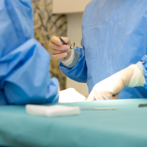
Sinus Laser Therapy (SiLaT) is a little invasive surgical technique involving the cleaning of the cyst wound to remove bacteria, hair, puss, and granulation tissue, which allows for tissue healing and, as a result, cyst cavity atresia.
This method is typically used for small lesions located in the upper part of the intergluteal cleft in patients who have not undergone other surgical procedures. In cases of recurrent cysts, open wounds, or lesions located near the anus, this method may not be effective.

This method is primarily used in cases of small lesions located in the upper part of the natal cleft, in patients who have not undergone any previous operations for pilonidal disease. In cases of recurrent pilonidal cysts, open wounds, or lesions located close to the anus, this method may not be sufficiently effective.
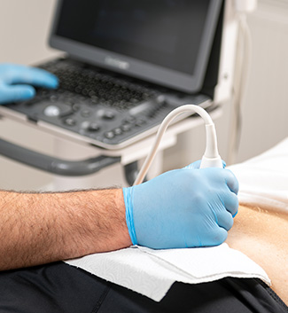
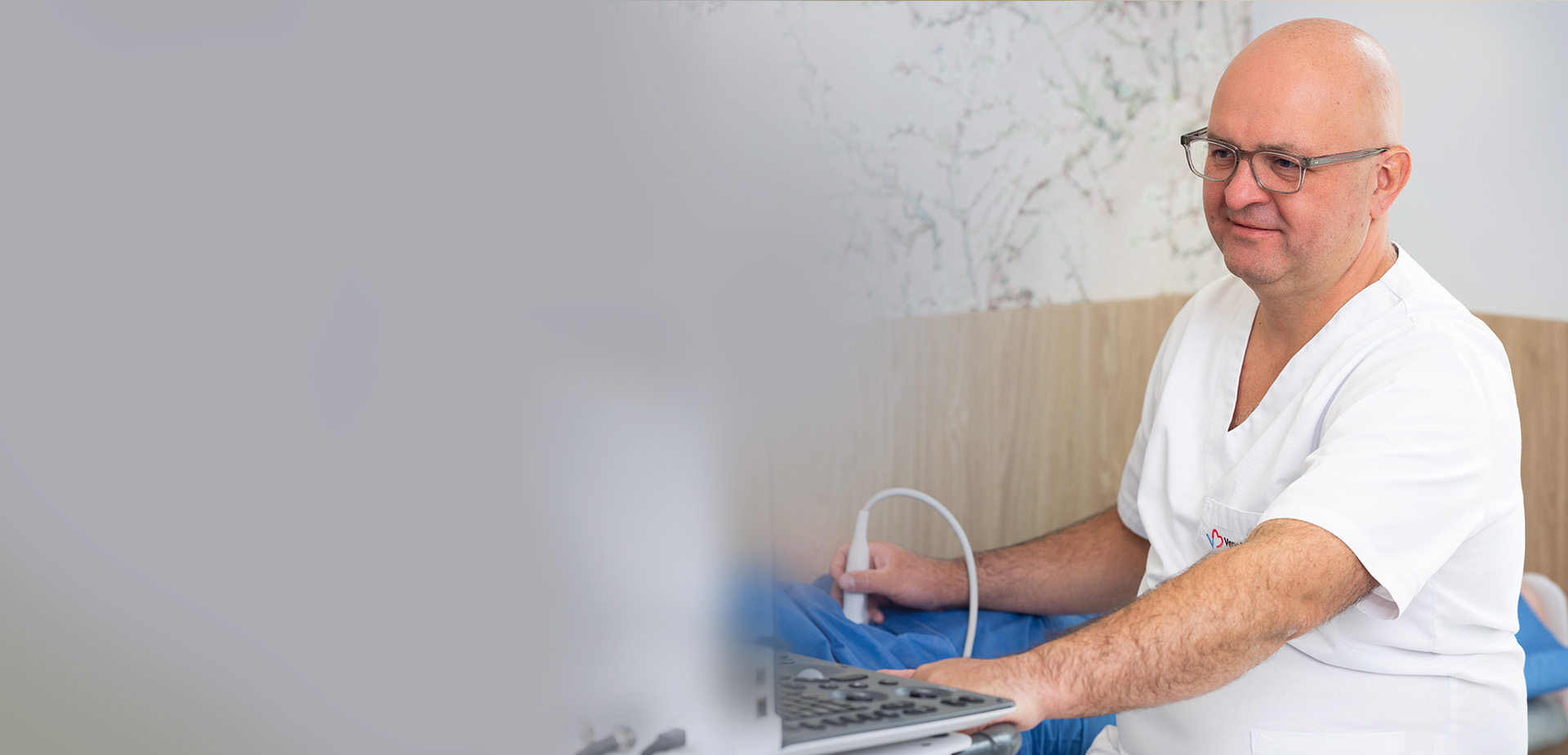
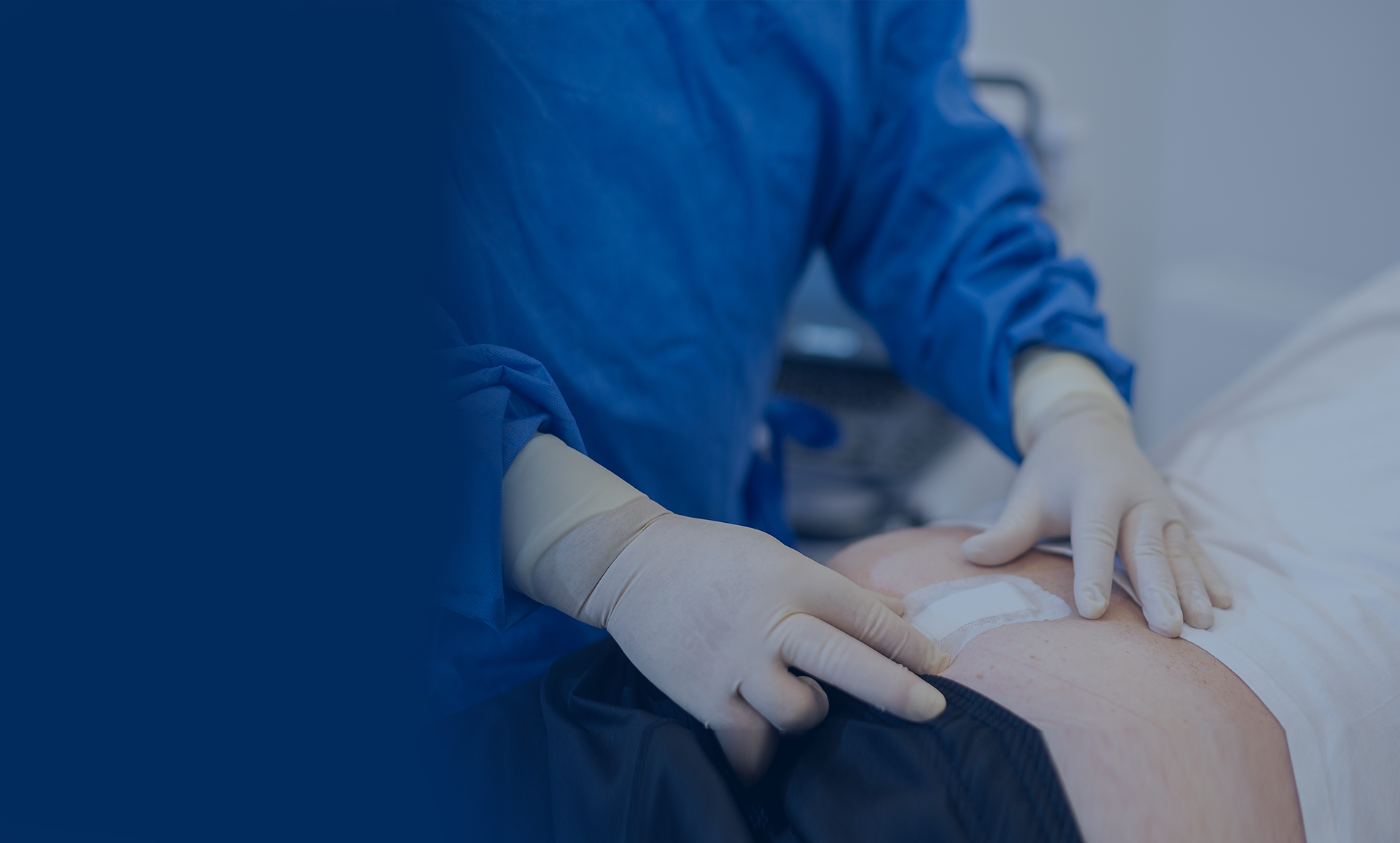
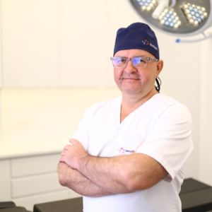
During the qualification visit, Dr. Norbert Zapotoczny assesses the lesion and establishes the final diagnosis based on the patient’s symptoms and the ultrasound. Then he discusses possible treatment methods with the patient. Each of the methods has its advantages and disadvantages, which is why discussing all the pros and cons with the patients is so important, since everyone may have different expectations and priorities regarding the treatment effects.
Book appointment
Laser treatment of pilonidal cyst is a little invasive procedure and, therefore, pain is usually experienced only on the first day after the procedure and is associated mostly with the use of the laser. The patient may walk and sit immediately after the procedure and 24 hours post surgery and take a shower without the dressing. Wound care is easy: the patient only needs to change the dressings, which can be done at home according to postsurgical instructions. The wound usually heals in 4 to 6 weeks. While normal physical activity is possible immediately after the procedure, intense exercise, contact sports, and activities associated with staying under water for long periods of time (bath, swimming pool) can be safely resumed 6 weeks after the procedure.
The success rate of laser ablation (SiLaT) in the treatment of pilonidal cysts is estimated at approximately 83–85% (over a 3-year follow-up period).
Due to the fact that the shape of the natal cleft remains unchanged, the risk of recurrence may increase over time.
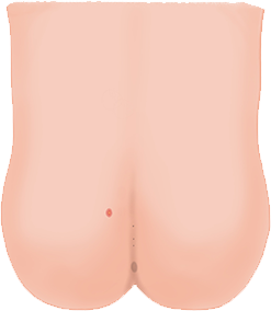
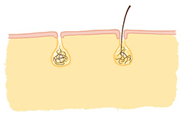
Comparison of treatment methodsA pilonidal cyst is a chronic subcutaneous abscess located at the bottom of the intergluteal cleft. A characteristic feature is the presence of hair within the sinus cavity, though this is not always present.
Typical symptoms of a pilonidal cyst include (not all may be present simultaneously):
Learn more about pilonidal cysts:
Several theories explain the formation of pilonidal sinuses. The most likely involves skin damage at the bottom of a tight, deep intergluteal cleft due to sweat and mechanical abrasion. This leads to secondary infection of superficial wounds, followed by bacterial penetration into the subcutaneous tissue, resulting in an abscess cavity connected to the skin surface via one or more fistulous tracts.
Despite the common theory of ingrown hairs, studies indicate that hairs found in pilonidal sinuses are loose hairs detached from the skin and trapped in the intergluteal cleft, often originating from the scalp. Many are short, stiff hairs with sharp ends, such as those cut at a barbershop, which penetrate the sinus through damaged skin or fistulas.
If the issue is recent and you’re awaiting a doctor’s visit, you can try simple methods to feel better at home before receiving professional care. These are detailed on our blog:
History of a patient with a pilonidal cyst
Pilonidal cyst – where does it come from and how can you treat it?
Why does the wound after Bascom Cleft Liftheal faster than after SiLaT?
Learn more about the condition and available treatments
Learn more